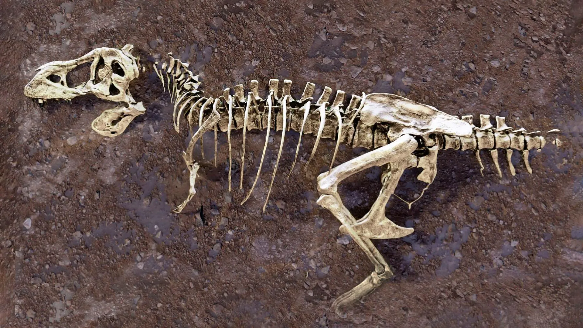
When paleontologists first recovered the massive fossilized rib from what is now recognized as the world's largest Tyrannosaurus rex, the bone presented a puzzle. A visible fracture suggested trauma, but the internal structure remained locked behind solid rock and mineralized tissue, inaccessible without risking damage to the rare specimen.
Researchers at the Department of Energy's Oak Ridge National Laboratory applied neutron imaging to address this limitation. The technique uses neutrons' ability to penetrate dense materials while interacting differently with various elements, allowing the construction of detailed three-dimensional images from within the fossil. The resulting scans revealed internal microstructures within the rib that have remained unseen since the dinosaur's death 66 million years ago.
The neutron images showed that the fracture observed on the exterior was accompanied by internal remodeling — a biological response indicating the animal survived the injury rather than dying immediately from it. This finding provides the first direct evidence of healing in a T. rex fossil, suggesting the animal lived for some time after sustaining the broken rib.
"The internal structure tells a story that the exterior alone cannot," said the lead scientist on the project, whose team published the findings this week. "We can see how the bone was trying to repair itself, which changes our understanding of how these animals dealt with injury."
The technology also revealed dense deposits within the bone cavity that correspond to what researchers describe as unusual mineralization patterns. These patterns differ from those typically observed in other T. rex specimens and may provide new data about the animal's growth rate, metabolism, or environment during its life.
Importantly, the neutron imaging approach preserved the integrity of the fossil. Traditional methods examining internal structures often require removing surrounding matrix or taking small samples, techniques that are not feasible with complete, rare specimens. The non-destructive nature of the neutron technique means the fossil remains available for future study while delivering its current insights.
The specimen, whose broken rib was the entry point for this investigation, represents the largest known Tyrannosaurus rex based on skeletal measurements. Its size has been a subject of scientific interest since its discovery, and the new internal data adds another layer to the understanding of how such massive animals functioned biologically.
The research team emphasized that the findings, while significant, represent a single specimen. They cautioned against drawing broad conclusions about all T. rex biology from one fossil, but noted that the methodology opens new pathways for studying other rare dinosaur fossils previously inaccessible to internal examination.
The publication of these findings marks a technical advancement as much as a biological one. By demonstrating that neutron imaging can successfully penetrate and image dense fossilized bone, the study provides a new tool for paleontologists worldwide. Future applications may include examining other specimens for signs of disease, injury, or growth patterns without risking damage to the remains.
As the research community processes these results, the broken rib of the world's largest T. rex stands as a testament both to the animal's resilience and to the evolving capabilities of scientific technology. The 66-million-year-old secret hidden within that bone is now accessible, not through force, but through the careful application of neutron science.
The study was published in a peer-reviewed geoscience journal and was supported by funding from the Department of Energy's Office of Science. Additional contributors included researchers from university paleontology departments and the neutron science division at Oak Ridge National Laboratory.